
CDVet e Ospedale Veterinario San Francesco presentano un caso clinico di Epatite Lobulare Dissecante (LDH) al Congresso Internazionale "Small Animal Hepatology" che si svolge a Venezia dal 30 marzo al 01 Aprile 2023.
.png)
CDVet e Ospedale Veterinario San Francesco presentano al Congresso Internazionale "Small Animal Hepatology" un caso clinico con aspetti di diagnostica per immagini e istopatologici, di Epatite Lobulare Dissecante (LDH), una rara patologia epatica del cane giovane.
Il caso clinico è stato oggetto di studio da parte del Dott. Yari Nebel (DVM - Ospedale Veterinario San Francesco) e del Dott. Riccardo Ferriani (DVM - Ospedale Veterinario San Francesco, Laboratorio Cdvet) e per la parte clinica e la Dott.ssa Alice Reginato (DVM, PhD, Dipl. ECVP - Laboratorio CDVET) per la parte Istolpatogica.
INTRODUCION
Lobular dissecting hepatitis (LDH) is formally defined as a chronic hepatitis characterized by a disruption of the normal lobular architecture due to the development of fibrosis. Even though a genetic predisposition is suspected, the etiology of LDH is largely unknown. Notwithstanding, data on etiology, pathogenesis, clinical and pathological presentation are largely lacking.
CASE DESCRIPTION
A 7-month-old, female mix-breed dog weighing 10kg with a body condition score of 3/9 presented for acute abdominal distension, sensory depression, melena and anorexia. On physical examination, abdominal enlargement with a fluid wave was noted, consistent with abdominal effusion. The dog was hospitalized, and hematology, serum biochemistry, serum TLI, folate, cobalamin, basal cortisol, coagulation panel and fecal flotation tests were run. The results were compatible with signs of hepatic damage associated with hepatic dysfunction. Abdominal ultrasound showed severe effusion associated with a reduction in portal flow, in the absence of specific hepatic parenchymal changes. The abdominal effusion was consistent with a low protein transudate (total solids 0,4g/dl); portal hypertension of hepatic origin was suspected.
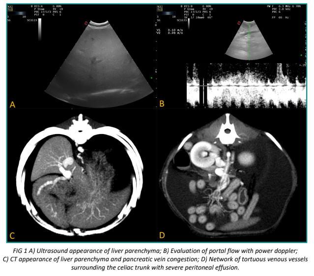
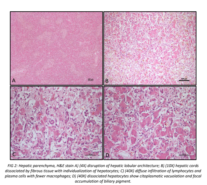
Differential diagnosis included LDH, arteriovenous fistula, portal vein system hypoplasia and occlusion of the caudal vena cava. Once the patient was deemed hemodynamically stable, computer tomography (CT) was performed, confirming portal hypertension, without evidence of parenchymal liver changes. A liver biopsy was proposed, however the owner elected euthanasia 72h after hospitalization due to rapid clinical deterioration. Liver samples were obtained post-mortem and submitted for histopathological characterization, which showed LDH with hepatocellular dissociation and individualization, dissecting fibrosis, necrosis and subacute lymphoplasmacytic and macrophagic infiltration. Additional histological analysis was performed on other organs, including the intestine, with no significant lesions noted.
DISCUSSION
LDH is rarely reported in veterinary medicine and only few complete case reports or series are available. It is believed that LHD occurs in young patients and is characterized by acute onset ascites, with change compatible with portal hypertension and hypoalbuminemia. The histological examination of the liver parenchyma (by biopsy or necropsy) is required for the diagnosis of LDH as imaging is often aspecific. However, the rapid progression of the disease hinders in-vivo clinical confirmation, so that the prevalence of the disease could be easily underestimated. Diagnostic imaging may similarly not be helpful in diagnosing the disease and may show aspecific patterns, as peculiarly described in this case. In the only available case report in which the CT was performed, the pattern was consistent with multiple hyperechoic nodules. Additionally, as the prognosis of LDH is poor, it may not allow to achieve diagnostic certainty due to the rapid and severe clinical worsening, leading to underestimation of the true prevalence of this primary liver disease. In conclusion, LDH should always considered as differential diagnosis when a young dog presents with acute and worsening signs of portal hypertension due to liver disease.
References
- Webster CRL, Center SA, Cullen JM, et al. ACVIM consensus statement on the diagnosis and treatment of chronic hepatitis in dogs. J Vet Intern Med. 2019;33(3):1173-1200.
- Mizooku H, Kagawa Y, Matsuda K, Okamoto M, Taniyama H. Histological and immunohistochemical evaluations of lobular dissecting hepatitis in American cocker spaniel dogs. J Vet Med Sci. 2013;75(5):597-603.
- Poldervaart JH, Favier RP, Penning LC, van den Ingh TSGAM, Rothuizen J. Primary hepatitis in dogs: a retrospective review (2002-2006). J Vet Intern Med. 2009;23(1):72-80.
- Buob S, Johnston AN, Webster CRL. Portal hypertension: pathophysiology, diagnosis, and treatment. J Vet Intern Med. 2011;25(2):169-186.
- Hostnik ET, Parker VJ, Cullen JM. Computed Tomography of Lobular Dissecting Hepatitis in a Young Golden Retriever. J Am Anim Hosp Assoc. 2019;55(2):E552-E03.